Heart Defects Glossary
Aortic Valve Stenosis
The aortic valve is narrow - the left ventricle has to work harder to pump the blood to the rest of the body. The more narrow the valve the harder the ventricle has to work.
Atrial Septal Defect (ASD)
A hole in the wall (septum) that divides the top two chambers of the heart (atrium).
Atrioventricular Canal (AV Canal)
A hole in the area where all four chambers meet. This includes the mitral and tricuspid valves. This defect ranges from a small opening between the atria (primum ASD) to a large hole in the center of the heart that causes an opening between the ventricles as well as between the atria. Sometimes the mitral and tricuspid valves become joined into a "common valve" that is insufficient and leaks.
Coarctation of the Aorta
A localized narrowing of the aorta just after it gives off the vessels to the head, neck and arms.
Dextrocardia
A peculiar condition in which the heart is positioned on the right side of the chest while it is normally on the left (mirror-image). The name is derived from dexter in Latin meaning "on the right" and cardio meaning "of the heart". If the rest of the organ systems are reversed, the condition is called situs inversus.

Double Outlet Right Ventricle
A very rare congenital heart defect in which both the pulmonary artery and the aorta, known as the great vessels, arise from the right ventricle. Normally, only the pulmonary artery that carries blood to the lungs for oxygen arises from the right ventricle. The aorta, which carries oxygenated blood from the heart to the body, normally arises from the left ventricle.
This defect almost always coincides with a ventricular septal defect (VSD), an abnormal opening in the septum (wall that separates the two ventricles) that allows blood to pass between the right and left ventricles. In the case of DORV, a ventricular septal defect is helpful because it allows the oxygen rich blood of the left ventricle passage to the right ventricle, which pumps it to the aorta and out of the heart to the body. If this passage didn’t exist, there would be no way for oxygen-rich blood to get to the aorta. Still, this oxygen rich blood mixes with oxygen poor blood, so oxygen levels in the blood are not optimal, and the heart must work extra hard to maintain circulation.
Ebstein’s Anomaly
Malformation of the right-sided tricuspid valve in which the valve is abnormally low in the right ventricle. This leads to an enlarged right atrium and a "leaky" tricuspid valve.
Hypoplastic Right Heart Syndrome
A defect that refers to the underdevelopment of the structures on the right side of the heart.
Problems present will include:
- Pulmonary valve atresia - (absence)
- Very small (or hypoplastic) right ventricle
- A small tricuspid valve
- A small (hypoplastic) pulmonary artery
Hypoplastic Left Heart Syndrome
Several cardiac defects that all involve underdevelopment of the left side of the heart. The structures typically effected are the left ventricle and the aorta. The left-sided valves (aortic and mitral) may be completely closed, meaning all of the blood must be pumped by the right ventricle alone.
Mitral Valve Stenosis
Narrowing of the mitral valve. If the narrowing is significant it can cause heart failure. If it is completely closed it is HLHS.
Patent Ductus Arteriosis (PDA)
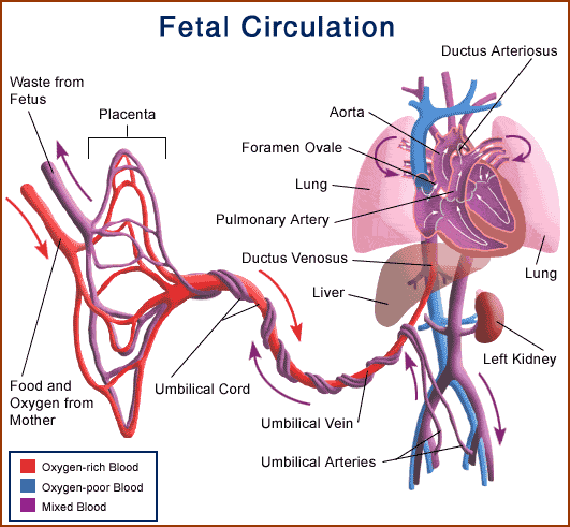
A condition caused by the failure of the ductus arteriosus to close. The ductus arteriosis normally is a temporary blood vessel connecting the pulmonary artery to the aorta that closes shortly after birth.
Patent Foramen Ovale
A defect in the atrial septum. It is an incomplete closure of the wall that results in the creation of a flap or a valve-like opening in the atrial septal wall. When a person with this defect creates pressure in their chest (sneezing, coughing,) the flap can open and cause oxygenated blood to mix with unoxygenated blood.
Pulmonary Valve Stenosis
Narrowing of the pulmonary valve. If the valve is completely closed the condition is called valve atresia.
Total Anomalous Pulmonary Venous Return (TAPV) or (TAPVR)
Rather than the blood supply returning from the lungs to the left atrium via the four distinct pulmonary veins as it should in a normal heart, the blood drains abnormally into the right atrium through a common pulmonary vein.
Shone’s Complex
A very rare defect that involves an abnormal mitral heart valve, a supramitral ring (membrane that covers the mitral valve) a subaortic stenosis (narrowing below the aortic valve) and coarctation of the aorta
Tetralogy of Fallot
A cardiac defect made up of four components:
- Narrowing of the pulmonary valve
- A large VSD
- A dilated aorta that is connected to both the right and the left ventricles
- Hypertrophy (thickening) of the right ventricle (This thickening results from the right ventricle having to work harder to pass blood across the narrowed pulmonary valve and into the lungs.
Transposition of the Great Arteries (TGA) / Transposition of the Great Vessels (TGV)
A reversal of the pulmonary artery and the aorta, which results in abnormal blood flow. In TGA rather than the blood flowing from the right side of the heart to the lungs to the left side of the heart throughout the body and back to the right side of the heart - the blood in a child with TGA flows from the right side of the heart to the body and back to the right side of the heart. Simultaneously, blood flows from the left side of the heart to the lungs and back to the left side of the heart.
Tricuspid Atresia
A defect where the tricuspid valve, which is normally located between the right atrium and the right ventricle, does not develop properly during pregnancy.
Basically, four problems are present:
- Improper development of the tricuspid valve prevents oxygen-poor (blue) blood from passing from the right atrium to the right ventricle and on to the lungs.
- The right ventricle is underdeveloped.
- VSD and ASD are present - allowing the blood to mix and be circulated
- PDA remains open allowing the blood to mix.
Tricuspid Valve Stenosis
Narrowing of the tricuspid valve. If the tricuspid valve is very small, the right ventricle is likely to be very small, and there may be no pulmonary valve opening.
Ventricular Septal Defect (VSD)
A hole or opening between the right and left ventricles. |


